
Prices in Germany are normally 75% below the cost for a spine test in the US and are individually quoted.
MRI

Magnetic Resonance Imaging (MRI), formerly referred to as Magnetic Resonance Tomography (MRT) or Nuclear Magnetic Resonance (NMR), is a method used to visualize the inside of living organisms as well as to detect the composition of geological structures. It is primarily used to demonstrate pathological or other physiological alterations of living tissues and is a commonly used form of medical imaging. The devices used in medicine are expensive, costing approximately $1 million USD per Tesla for each unit (common field strength ranges from 0.3 to 3 Teslas), with several hundred thousand dollars per year for maintenance. The stronger the field the greater the resolution.
MRIs are particularly useful in diagnosis of Degenerative Disc Disease (it detects the loss of water in the disc nucleus), Spinal Stenosis or a herniated disc. In short, MRIs are best suited for soft tissue examination. For hard tissue (bones) examination the CAT scan provide more information.
CAT Scan
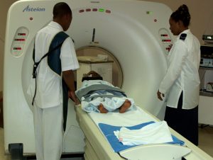
Computed tomography (CT), originally known as computed axial tomography (CAT or CT scan) and body section roentgenography, is a medical imaging method employing tomography where digital geometry processing is used to generate a three-dimensional image of the internals of an object from a large series of two-dimensional X-ray images taken around a single axis of rotation. The word "tomography" is derived from the Greek tomos (slice) and graphia (describing). CT produces a volume of data which can be manipulated, through a process known as windowing, in order to demonstrate various structures based on their ability to block the x-ray beam. Although historically (see below) the images generated were in the axial or transverse plane (orthogonal to the long axis of the body), modern scanners allow this volume of data to be reformatted in various planes or even as volumetric (3D) representations of structures.
Bone Scan
A bone scan comes under the auspices of nuclear medicine. The patient injected with a small amount of radioactive material and then scanned with a Gamma camera, a device sensitive to the radiation emitted by the injected material.
Dexa bone scans are recommended for assessing bone density. The expose the patient to little radiation and often more accurate than other methods.
Discogram
A discogram is a fluoroscop (X-ray TV) examination of the disc. The test is performed under local anethesia by injecting dye into the center of the suspected injured disc(s). The dye makes the disc clearly visible on X-ray film and a fluoroscope screen.
A discogram reveals damage to a disc and if those discs cause pain. Details from the discogram can show tears to the annulus, the outer covering of the disc. During the procedure, fluid is injected into the disc to increase the pressure inside the disc to check for leakage. If the patient experiences pain during the procedure, there is a good chance the disc is diseased.
A discogram is usuallly done if an MRI fails to show a herniated disc and surgery is being seriously considered. The discogram reveals the nucleus of the disc, not the surrounding tissue.
Electromygram
An electromyogram (EMG) test looks for abnormal electrical signaling patterns in muscles. A pinched nerve in any area of the body will act differently than a nerve under no pressure. Consequently the muscles they control will act abnomally. It is the difference in patterns (between normal and abnormal) that can lead to isolating the nerves affected by wayward disc or spinal stenosis.
Myelography
Myelography is a type of radiographic examination which uses a contrast medium to detect pathology of the spinal cord, including the location of a spinal cord injury, cysts, and tumors. The procedure often involves injection of contrast medium into the cervical or lumbar spine, followed by several X-ray projections.
Myelography has been largely replaced by the use of CT and MRI scans.