
The gastrointestinal tract or digestive tract, also referred to as the GI tract or the alimentary canal (nourishment canal) or the gut, is the system of organs within multicellular animals which takes in food, digests it to extract energy and nutrients, and expels the remaining waste. This process is called digestion.
Basic anatomy
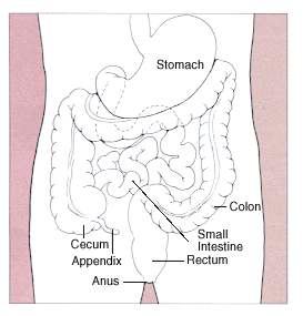
In a normal human adult male, the GI tract is approximately 7.5 meters long (25 feet) and consists of the following components:
Upper Gastrointestinal Tract
- Mouth (buccal cavity; includes salivary glands, mucosa, teeth and tongue)
- Pharynx
- Oesophagus or Esophagus (gullet) and cardia
- Stomach, which includes the antrum and pylorus and pyloric sphincter.
Lower gastrointestinal tract
The small intestine has three parts:
- duodenum
- jejunum
- ileum
The large intestine, which has three parts:
- caecum (the vermiform appendix is attached to the cecum)
- colon (ascending colon, transverse colon, descending colon and sigmoid flexure)
- rectum
Related organs
The liver secretes bile into the small intestine via the biliary system, employing the gallbladder as a reservoir. The pancreas secretes an isosmotic fluid containing bicarbonate and several enzymes, including trypsin, chymotrypsin, lipase, and pancreatic amylase, as well as nucleolytic enzymes (deoxyribonuclease and ribonuclease), into the small intestine. Both these secretory organs aid in digestion.
Physiology
Specialization of Organs
Four organs are subject to specialization in the kingdom Animalia.
The first organ is the tongue which is only present in the phylum Chordata.
The second organ is the esophagus. The crop is an enlargement of the esophagus in birds, insects and other invertebrates that is used to store food temporarily.
The third organ is the stomach. In addition to a glandular stomach (proventriculus), birds have a muscular "stomach" called the ventriculus or "gizzard." The gizzard is used to mechanically grind up food.
The fourth organ is the large intestine. An outpouching of the large intestine called the cecum is present in non-ruminant herbivores such as rabbits. It aids in digestion of plant material such as cellulose.
Immune function
The gastrointestinal tract is also a prominent part of the immune system (Coico et al 2003). The low pH (ranging from 1 to 4) of the stomach is fatal for many microorganisms that enter it. Similarly, mucus (containing IgA antibodies) neutralizes many of these microorganisms. Other factors in the GI tract help with immune function as well, including enzymes in the saliva and bile. Health enhancing intestinal bacteria serve to prevent the overgrowth of potentially harmful bacteria in the gut.
Histology
The GI tract has a uniform general histology with some differences which reflect the specialization in functional anatomy. The GI tract can be divided into 4 concentric layers:
- mucosa
- submucosa
- muscularis externa (the external muscle layer)
- adventitia or serosa
Mucosa
The mucosa is the innermost layer of the GI tract, surrounding the lumen, or space within the tube. This layer comes in direct contact with the food (or bolus), and is responsible for absorption and secretion, important processes in digestion.
The mucosa can be divided into:
- epithelium
- lamina propria
- muscularis mucosae
The mucosa are highly specialized in each organ of the GI tract, facing a low pH in the stomach, absorbing a multitude of different substances in the small intestine, and also absorbing specific quantities of water in the large intestine. Reflecting the varying needs of these organs, the structure of the mucosa can consist of invaginations of secretory glands (i.e. gastric pits), or it can be folded in order to increase surface area (i.e. villi, microvilli).
Submucosa
The submucosa consists of a dense irregular layer of connective tissue with large blood vessels, lymphatics and nerves branching into the mucosa and muscularis. It contains Meissner's plexus, an enteric nervous plexa, situated on the inner surface of the muscularis externa.
Muscularis externa
The muscularis externa consists of a circular inner muscular layer and a longitudinal outer muscular layer. The circular muscle layer prevents the food from going backwards and the longitudinal layer shortens the tract. The coordinated contractions of these layers is called peristalsis and propels the bolus, or balled-up food, through the GI tract. Between the two muscle layers are the myenteric or Auerbach's plexa.
Adventitia/Serosa
The adventitia consists of several layers of connective tissue. When the adventitia is facing the mesentery or peritoneal fold, the adventitia is covered by a mesothelium supported by a thin connective tissue layer, together forming a serosa, or serous membrane.